Broken Ankle: Causes, Symptoms, and Treatment
A broken ankle occurs when one or more of the bones forming the ankle joint are fractured, leading to pain, swelling, and difficulty bearing weight. The injury can result from minor incidents such as a misstep or sports activity, or from more severe trauma like a fall or road accident. Timely assessment and treatment by an orthopedic specialist can help support proper recovery and joint stability.
In line with the Singapore Ministry of Health (MOH) guidelines, early medical attention and appropriate management are important to reduce the risk of long-term complications. This guide outlines the causes, symptoms, and treatment options for a broken ankle to provide a clearer understanding of the condition.”
Also known as an ankle “fracture”, a broken ankle means that one or more of the bones that form the ankle joint are broken.
Fractures can have varying degrees of severity. It can range from a simple break in a bone, where one might still be able to walk, to several fractures which can render your ankle out of place and would require one to prevent putting weight on it for months.
Basically, it means that the more broken bones there are, the more unstable the ankle will be. The ligaments, which hold the joint and ankle bones in position, may also be damaged.
People of all ages can be affected by broken ankles. In the past 30 to 40 years, experts have observed an increase in the cases and severity of fractures. This was said to be partly due to an older and more active, older population of baby boomers. Hence, it is advisable to visit an orthopaedic clinic in Singapore for an accurate diagnosis and the right treatment as soon as possible.
In this article, we will cover what causes foot and ankle fractures and which parts of your foot are affected. We will also cover the various symptoms behind foot and ankle fractures and the many ways they are examined and treated. Ankle fractures in Singapore are treated in different ways by foot doctors or ankle specialists, depending on the type and severity of the condition.
What is a Broken Ankle?

The ankle joint is made up of three bones:
- Tibia, the shinbone
- Fibula, a smaller bone of the lower leg
- Talus, a small bone sitting between the heel bone (calcaneus), the tibia and fibula
There are specific parts of the tibia and fibula that form the ankle:
- Medial malleolus, the inside part of the tibia
- Posterior malleolus, the back part of the tibia
- Lateral malleolus, the end of the fibula
Ankle fractures are classified based on whichever area of the bone is broken. For example, if the fracture is at the end of the fibula, it is called a lateral malleolus fracture. If both the fibula and tibia are broken, it will be referred to as a bimalleolar fracture.
There are two joints involved in fractures:
- Ankle joint, where the talus, fibula and tibia meet
- Syndesmosis joint, in the middle of the fibula and tibia, held together by ligaments
The ankle joint is made stable by multiple ligaments.
What Causes a Broken Ankle?
Ankle fractures are a common type of traumatic injury to the foot in Singapore and can occur due to various reasons, ranging from daily activities such as running and playing sports to unforeseen circumstances like a car accident, especially since we use our ankles so much on a daily basis.

- Direct impact to the ankle e.g. from a car accident
- Rolling of one’s ankle
- Falling down or tripping
- Rotating or twisting of one’s ankle
What are the symptoms of a broken ankle?
Every foot or ankle injury should be evaluated by a doctor in Singapore as pain from an ankle fracture can feel like an ankle sprain. Sometimes patients with flatfeet are more prone to injury or twisting their ankle. In the case of an ankle fracture, not getting treatment early may result in a nonunion or delayed union in which the bone does not heal; causing swelling, tenderness and pain that becomes worse over time. Immediate treatment is required to help you regain mobility and strength to complete daily activities and get back to doing what you love. It is advisable to never ignore the symptoms and to seek medical attention as soon as possible.

Every foot or ankle injury should be evaluated by a doctor. This is because a broken ankle can feel like a serious ankle sprain.
Some usual symptoms of a broken foot or ankle are:
- A sharp and immediate pain
- Bruising
- Swelling
- Tenderness
- Unable to place any weight on the injured foot
- Difficulty in walking or of movement around the area
- Blisters
- Foot or ankle might look out of place, especially if the ankle joint is also dislocated
- Bones protruding through the skin (in severe cases)
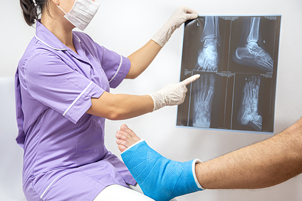
How is a Broken Ankle Diagnosed?

A physical examination and a look into the patient’s medical history
First, the doctor will discuss the patient’s medical history, symptoms and how the injury occurred. Then, a close examination of the ankle, foot and lower leg will be done.
Imaging tests
If a fracture is suspected, the doctor will use additional tests to gain more insight into the injury.
X-rays
The most common and widely used diagnostic imaging technique are X-rays. They are able to show whether the bone is broken and if there is any displacement or gap between the broken bones. One can also tell how many pieces of broken bones there are. Additionally, X-rays of the leg, ankle and foot may be taken to ensure that nothing else is injured.
Stress test
Based on the kind of fracture, the doctor may apply pressure on the ankle and take a special X-ray. This is known as a stress test, which is done to see if certain of the fractures require any surgery.
Computed tomography (CT) scan
A CT scan is able to create a cross-section image of the ankle. It is conducted to further evaluate the injury. This is particularly useful at times when the fracture extends into the ankle joint.
Magnetic resonance imaging (MRI) scan
MRI scans can produce high-resolution images of both bones and soft tissues, such as ligaments. An MRI scan may be conducted to evaluate the ankle ligaments for certain fractures.
Broken Ankle Treatment Options
How is a lateral malleolus fracture treated?
A lateral malleolus fracture refers to a fracture of the fibula. The fibula can be fractured at several different levels. The level of the fracture may direct how the treatment is conducted.
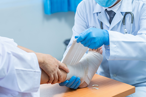
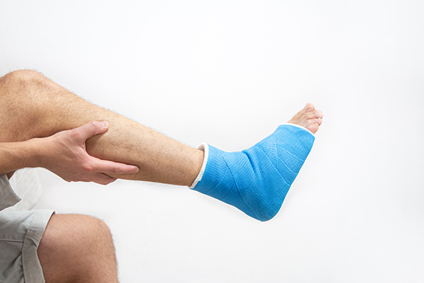
Nonsurgical treatment
To see if the ankle is stable, a stress x-ray may be conducted. If the ankle is stable and the broken bone is barely out of place, surgery may not be required. The part of the bone that is broken may also determine the kind of treatment required.
There are various methods used to protect the fracture while it heals. They range from a high-top tennis shoe to a short leg cast. Certain physicians may allow their patients to apply weight on their leg immediately, whereas others might require them to wait for up to 6 weeks.
To ensure that fragments of the fracture have not moved out of place during the healing process, patients have to visit the doctor regularly to repeat their x-rays.
Surgical treatment
In the event that the fracture is out of place or the joint is unstable, the need for surgery for a broken ankle may arise. In this procedure, the fragments of the bone are repositioned into their normal alignment, then held in place using special screws and metal plates attached to the outer surface of the bone. In certain cases, to keep the bone fragments in place while they heal, a rod or screw may be used inside the bone.
How is a medial malleolus fracture treated?

A medial malleolus fracture refers to a break in the tibia, located at the inside of the lower leg. Fractures can occur at various levels of the medial malleolus. Such fractures usually occur along with a lateral malleolus (fracture of the fibula), a posterior malleolus (fracture of the back of the tibia), or an injury to the ankle ligaments.
Nonsurgical treatment
The fracture need not be treated with surgery if it is not out of place, or if it is a very low one with very small pieces. To determine if the fracture and ankle are stable, a stress x-ray may be conducted.
A short leg cast or removable brace may be used to treat the fracture. Typically, one would need to refrain from applying weight on their leg for about 6 weeks. Frequent visits to the doctor for repeat x-rays will be required to ensure the fracture does not move and stays in its position.
Surgical treatment
Surgery may be recommended if the ankle is unstable or if the fracture is out of position.
For certain cases, even if the fracture is in place, surgery may still be considered as an option. This is to reduce the risk of nonunion, where the fracture does not heal, and to allow the ankle to start moving earlier.
In a medial malleolus fracture, the ankle joint may be impacted or indented. Impaction occurs when a great force drives the end of one bone into another. To repair an impacted fracture, bone grafting may be required. The graft acts as a scaffolding for new bone to grow on and can lower the risk of arthritis developing later.
Additionally, based on the fracture, bone fragments may also be fixed using screws, a plate and screws, or various wiring methods.
How is a posterior malleolus fracture treated?
A posterior malleolus fracture refers to a fracture of the back of the tibia at the level of the ankle joint.
The fibula (lateral malleolus) is also broken in the majority of posterior malleolus fracture cases. This is due to the fact that it shares ligament attachments with the posterior malleolus. There is also a possibility of a fracture of the medial malleolus.
The back of the ankle may be unstable based on how big the broken piece of bone is. From certain studies, it is said that the ankle should be treated with surgery if the piece of broken bone is bigger than 25% of the ankle joint.
Due to the risk of arthritis developing, it is essential for a posterior malleolus fracture to be diagnosed and treated properly. The smooth surface that lines a joint is called cartilage. It covers the back of the tibia where the bone breaks. The cartilage will not heal properly if the broken piece of bone is larger than around 25% of the ankle and is more than a few millimetres out of place. As such, the surface of the joint will not be smooth. This leads to increased and uneven pressure on the joint surface, which eventually leads to cartilage damage and the development of arthritis.
Nonsurgical treatment
The fracture can be treated without surgery if it is stable and in place. A short leg cast or removable brace may be used, and patients will normally be advised to refrain from putting any weight on the ankle for 6 weeks.
Surgical treatment
Surgery may be recommended if the fracture is unstable or out of position.
There are various surgical options available for the treatment of posterior malleolar fractures. One method is to place screws from the front of the ankle to the back, or vice versa. Another method would be to place a plate and screws along the back of the shin bone.
How are bimalleolar fractures or bimalleolar equivalent fractures treated?

Since “bi” means two, “bimalleolar” means that two out of three parts or malleoli of the ankle are broken.
In the majority of bimalleolar fracture cases, the lateral malleolus and medial malleolus are broken and the ankle is not stable.
In a bimalleolar equivalent fracture, the ligaments on the inner (medial) side of the ankle are injured in addition to one of the malleoli being fractured. This typically means that the fibula is broken along with injury to the medial ligaments, causing the ankle to be unstable.
To determine whether the medial ligaments are injured, a stress test x-ray may be conducted.
Bimalleolar fractures or bimalleolar equivalent fractures are unstable fractures. They can be associated with a dislocation.
Nonsurgical treatment
Surgery is normally recommended for such injuries, which are considered unstable.
If the patient has significant health problems, where the risk of surgery may be too high, or if they do not usually walk, then nonsurgical treatment might be considered.
Normally, immediate treatment involves the use of a splint to immobilize the ankle until the swelling is reduced. A short leg cast is then applied, which may be changed frequently as the swelling in the ankle subsides.
Frequent doctor visits would be required to repeat x-rays and ensure the ankle remains stable.
Weight-bearing will not be allowed for 6 weeks in the majority of cases. After that, as the ankle continues to heal, it may be protected by a removable brace.
Surgical treatment
Most of the time, surgical treatment will be recommended for lateral and medial malleolus fractures as they make the ankle unstable. They are treated using the same surgical methods as the other fractures listed above.
How is a trimalleolar fracture treated?

Since “tri” means three, a trimalleolar fracture would mean that all three malleoli of the ankle are broken. These are unstable injuries and can even be associated with a dislocation.
Nonsurgical treatment
As these surgeries are considered unstable, surgery is typically recommended.
Just like bimalleolar fractures, nonsurgical treatment may be considered if the patient has significant health issues where the risk of surgery may be too high, or if they do not usually walk.
The nonsurgical treatment methods are similar to that of bimalleolar fractures.
Surgical treatment
Each fracture can be treated using the same surgical methods as the other fractures listed above.
How is a syndesmotic injury treated?

The syndesmosis joint is located between the tibia and fibula. It is held together by ligaments. A syndesmotic injury may just be to the ligament, also known as a high ankle sprain. Based on how unstable the ankle is, such injuries may be treated without the need for surgery. However, the healing process of these sprains is longer than a normal ankle sprain.
More often than not, a syndesmotic injury would include both a ligament sprain and one or more fractures. These are unstable injuries, and proper surgical treatment is highly recommended for stable recovery.
A stress test x-ray may be conducted by the doctor to determine whether the syndesmosis is indeed injured.
What does the recovery process of a broken ankle look like

Since there is a large variety of injuries, there are also varying degrees of how quickly people can heal from their injury. Typically, it takes at least 6 weeks for the broken bones to heal, but it may take even longer for the involved tendons and ligaments to heal.
As previously mentioned, repeated x-rays will likely be used to monitor bone healing. This is normally done more frequently during the first 6 weeks with nonsurgical treatments.
Pain management
After an injury or surgery, medications are often prescribed for short-term pain relief. There are various types of medicines available to help manage pain. These include opioids, non-steroidal anti-inflammatory drugs (NSAIDs) and local anaesthetics. A combination of these medications may be used to improve pain relief and also reduce the need for opioids.
Although opioids help to relieve pain after injuries and surgeries, they are narcotic and can be addictive. As such, one should be wary and only use them as directed by a doctor. Once the pain begins to improve, the use of opioids should be stopped. If the pain has not begun to improve within a few days of treatment, a doctor should be consulted.
Rehabilitation
Regardless of how a fracture is treated, rehabilitation is still crucial.
Once a physician allows for movement of the ankle to begin, it is important to engage regularly in physical therapy and home exercise programs.
Eventually, the patient will also have to do strengthening exercises. It may take several months for the muscles around the ankle to regain their strength. It is crucial to do these exercises regularly to get strong enough for returning to regular activities and walking without a limp.
Weight bearing
When one can start putting weight on their ankle is determined by the fracture itself. Once the physician feels that the injury is stable enough, they will allow the patient to start putting weight on their ankle.
It is crucial that no weight is put on the ankle until a physician allows it. If weight is put on the ankle too early, the fracture fragments may move or the surgery may fail and require a restart.
Supports
There are many supports that one can wear on their injured ankle, depending on the injury.
In the beginning, most ankle fractures are put in a splint to allow for protection of the ankle and for the swelling to go down. Afterwards, a cast or removable brace may be used.
The physician may also recommend wearing an ankle brace for several months while participating in sports, even after the fracture has healed.
What are the possible complications after fracture treatment?
The elderly, smokers or people with diabetes face a higher risk of complications after surgery, including problems with wound healing. This is due to the fact that their bones may take longer to heal.
Nonsurgical treatment
Without surgery, there is a risk of the fracture moving out of place before it is healed. As such, it is necessary to go for follow-up visits with a doctor as scheduled.
A malunion occurs when the fracture fragments move out of place and the bones heal in that position. Treatment is determined based on how the stability of the ankle joint is affected and how far out of place the bones have moved.
Arthritis can eventually be developed if a malunion occurs or if the ankle becomes unstable after it heals.
Surgical treatment
The general risks of surgical treatment are:
- Pain
- Bleeding
- Blood clots in the leg
- Infection
- Damage to blood vessels, tendons, or nerves
The risks of treating ankle fractures surgically are:
- Development of arthritis
- Difficulty with bone healing
- Pain caused by screws and plates used to fix the fracture. As such, some patients choose to remove them months after the fracture has healed.
What are the outcomes of fracture treatment?

The majority of patients are able to return to their normal non-intensive daily activities within 3 to 4 months. However, studies have shown that some people can still be recovering up to 2 years after their injuries. It may take several months for one to be able to walk without a limp and return to their previous competitive level in sports. With regards to driving, most people are able to resume within 9 to 12 weeks from the time of injury.

- Do I have weak bones?
- If surgery is required, what risks do I face?
- Am I at risk of not doing well from treatment?
- How long will I have to be on medical leave for?
- When will I be able to start putting weight on my leg?
Frequently Asked Questions
What are the common causes of a broken ankle?
A broken ankle can occur due to falls, twisting injuries, sports activities, or high-impact accidents. These incidents may cause one or more bones around the joint to fracture. Seeking timely assessment from an orthopaedic clinic in Singapore ensures proper diagnosis and management.
What symptoms should I look out for?
Typical symptoms include swelling, bruising, pain when bearing weight, and deformity around the joint. In severe cases, the foot may appear misaligned or unstable.
How is a broken ankle diagnosed?
Doctors typically use physical examination and imaging tests such as X-rays or CT scans to confirm the type and severity of the fracture. A consultation with an ankle specialist in Singapore can help determine a suitable treatment plan.
What treatment options are available?
Treatment depends on the fracture’s stability and alignment. Stable fractures may only require immobilisation with a cast or boot, while displaced fractures may need surgical repair.
How long does recovery from a broken ankle take?
Recovery duration varies by injury severity. Mild fractures may heal within six weeks, while complex fractures can take several months. Physical therapy is often recommended to restore movement and strength.
When should I seek medical attention?
If you experience severe pain, swelling, or difficulty walking after an injury, seek prompt evaluation at our Mount Elizabeth Novena orthopaedic clinic. Early intervention can help prevent complications and promote proper healing.