Everything You Need To Know About ACL Reconstruction Surgery

This article provides detailed information on anterior cruciate ligament (ACL) injuries. The following discusses ACL anatomy and the pathophysiology of an ACL tear, as well as treatment options for ACL injuries. ACL surgery techniques and rehabilitation, potential complications, and outcomes are also included. This page is intended to assist the patient in making an informed decision about how to manage an ACL injury.
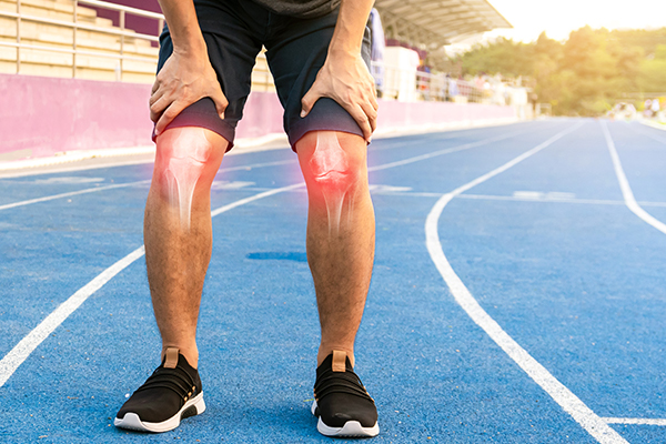
What is an ACL tear or injury?

The anterior cruciate ligament (ACL), which is the dense connective tissue that runs through the knee and connects the femur (thigh bone) to the tibia (shin bone), is the most essential of the four ligaments that connect the two. It is one of the most injured ligaments in the knee because it provides structural support during strenuous activity. People who participate in high-risk activities such as football, soccer, basketball, and skiing are more likely to sustain ACL tears.
In essence, the knee is a hinged joint that is held together by the medial collateral (MCL), lateral collateral (LCL), anterior cruciate (ACL) and posterior cruciate (PCL) ligaments. The ACL runs diagonally in the centre of the knee, preventing the tibia from sliding out in front of the femur, along with providing rotational stability to the knee. A layer of articular cartilage covers the weight-bearing surface of the knee. The medial meniscus and lateral meniscus are on either side of the joint, between the cartilage surfaces of the femur and tibia. These menisci function as shock absorbers and work with the cartilage to reduce the stresses between the tibia and femur. That’s why when an individual sustains an ACL tear, they will have difficulty using their knee down to the foot.
About half of all ACL tears occur along with damage to the meniscus, articular cartilage, or other ligaments. In addition, patients may have bruises of the bone beneath the cartilage surface. These bruises may be detected on a magnetic resonance imaging (MRI) scan and may indicate injury to the overlying articular cartilage. In more severe cases, treatment may involve ACL surgery to restore stability and function.
Symptoms of an ACL Tear
The symptoms of an ACL tear can vary depending on the severity of the injury. While some patients experience immediate discomfort, others may notice instability or functional limitations developing over time.
A Popping Sensation
Some individuals report hearing or feeling a “pop” in the knee at the time of injury. This sensation is often associated with a sudden ligament rupture, particularly during sports or abrupt movements.
Intense Knee Pain
Pain is commonly felt immediately after the injury. The level of discomfort can range from moderate to severe and may worsen with movement or weight-bearing.
Rapid Swelling
Swelling typically develops within a few hours due to bleeding within the knee joint. This swelling can cause tightness and restrict normal knee movement.
Knee Instability
A common symptom of an ACL tear is the feeling that the knee may “give way,” especially during turning, pivoting, or changing direction. This instability can affect confidence during movement. If this occurs, it is advisable to consult a knee specialist for a proper assessment and appropriate management.
Difficulty Bearing Weight
Some patients find it difficult to place weight on the affected leg, particularly in the early stages after injury. Weight-bearing may increase pain or create a sense of weakness in the knee.
Limited Range of Motion
Swelling, pain, and joint stiffness can limit the knee’s ability to fully bend or straighten. This restriction may persist without appropriate treatment or rehabilitation.
Difficulty Walking
Everyday activities such as walking, climbing stairs, squatting, or kneeling may become uncomfortable or unstable, affecting daily function and mobility.
What causes an ACL tear?

An ACL tear commonly occurs when excessive force or abnormal movement places stress on the knee joint. These injuries are frequently seen in sports and activities that involve rapid changes in movement, but they can also occur during everyday activities.
Sudden Deceleration or Stops
Abruptly slowing down or stopping while the foot is planted can cause the thigh bone to shift forward over the shin bone. This places strain on the ACL, increasing the risk of a tear.
Pivoting or Twisting Movements
Quick changes in direction, especially when pivoting on one leg, can cause rotational stress at the knee. This mechanism is a common cause of non-contact ACL tears in sports such as football, basketball, and netball.
Awkward Landings
Landing from a jump with poor knee alignment, limited knee flexion, or uneven weight distribution can overload the ligament. Repeated exposure to such landings raises the likelihood of ACL injury.
Hyperextension of the Knee
An ACL tear may occur when the knee is forced to straighten beyond its normal range of motion. This can happen during sports collisions or accidental slips and falls.
Direct Trauma
Although less common than non-contact mechanisms, a direct blow to the knee, such as during a collision with another player or object, can result in an ACL tear by forcing the joint into an unstable position.
How is an ACL tear diagnosed?

Diagnosing an ACL tear involves a combination of clinical assessment and imaging studies. This approach helps determine the extent of the injury and whether other structures within the knee joint are also affected.
Physical Examination (Clinical Assessment)
A session at our orthopaedic clinic at Mount Elizabeth, Novena usually begins with a detailed clinical evaluation. This allows our doctor to assess knee stability, movement, and functional limitations.
This may include:
Discussion of Medical History
The doctor will ask about how the injury occurred, the presence of symptoms such as swelling or instability, and whether a popping sensation was felt at the time of injury. Information about sports participation or previous knee injuries is also considered.
Movement Assessment
The knee is examined for swelling, range of motion, tenderness, and signs of instability. Comparisons are often made between the injured knee and the unaffected side.
Lachman Test
The Lachman test is commonly used to assess the integrity of the ACL. During this test, the examiner evaluates the amount of forward movement of the tibia relative to the femur. Increased movement and a soft endpoint may indicate an ACL tear.
Anterior Drawer Test and Pivot Shift Test
These tests further assess knee stability and ligament function. Abnormal movement during these tests may suggest damage to the ACL or other supporting ligaments of the knee.
Imaging Tests
Imaging studies are used to confirm the diagnosis and evaluate the extent of the injury.
MRI (Magnetic Resonance Imaging)
An MRI scan provides detailed images of the ACL and surrounding structures. It helps identify the severity of the tear and detect associated injuries to the meniscus, cartilage, or other ligaments.
X-ray
X-rays are often performed to rule out fractures or bone-related injuries. While X-rays do not show ligament damage, they help exclude other possible causes of knee pain following trauma.
What Are the Possible Treatments for an ACL Tear?

Treatment for an ACL tear depends on factors such as the severity of the injury, knee stability, activity level, age, and overall lifestyle needs. In Singapore, both non-surgical management and ACL surgery may be considered as part of an individualised treatment plan.
Non-surgical Treatment
Non-surgical treatment focuses on rehabilitation to restore knee strength, stability, and function. This approach typically involves structured physiotherapy, activity modification, and, in some cases, the use of a knee brace to reduce instability.
Nonsurgical management may be considered for:
- Individuals with partial ACL tears and no clinical signs of knee instability
- Patients with complete ACL tears who do not experience instability during daily activities or low-impact sports and are willing to avoid high-impact activities
- Those who lead a sedentary lifestyle or perform light manual work
- Children or adolescents whose growth plates are still open
While non-surgical treatment can be recommended for selected patients, some individuals may experience recurrent episodes of knee instability. This can increase the risk of secondary injuries to the meniscus or cartilage over time.
ACL Surgery as a Treatment Option
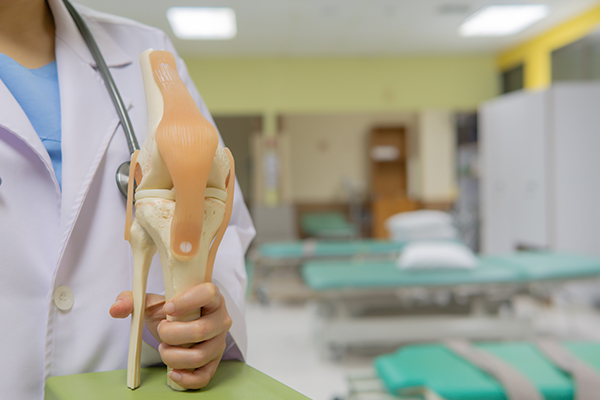
ACL reconstruction surgery is usually recommended for patients with persistent knee instability, combined ligament injuries, or functional demands that require a stable knee. Rather than repairing the torn ligament, the procedure involves reconstructing the ACL using a tendon graft.
The choice of graft depends on patient-specific factors, surgical considerations, and clinical judgement.
Patellar Tendon Autograft
This graft uses a portion of the patient’s own patellar tendon along with small bone plugs. It is often considered for physically active individuals. While it is designed to offer strong fixation, it may be associated with anterior knee pain or discomfort when kneeling in some patients.
Quadriceps Tendon Autograft
The quadriceps tendon autograft uses tissue from the front of the thigh and may be considered in selected cases, including revision surgeries. It provides a larger graft but may be associated with postoperative anterior knee discomfort and a longer recovery period for some patients.
Allograft (Donor Tissue)
Allografts use tendon tissue obtained from a donor. This option avoids graft harvesting from the patient and may result in shorter surgical time and smaller incisions. However, allografts may carry considerations such as graft incorporation time and suitability for younger or highly active individuals.
How is ACL Surgery Conducted?

ACL reconstruction surgery is typically performed using minimally invasive techniques. The procedure is carried out in stages, with preparation before surgery and structured steps during and after the operation to support recovery.
1. Preparation and Anaesthesia
Before surgery, patients often undergo a period of physiotherapy to reduce knee swelling and restore range of motion. Performing ACL surgery on a stiff or swollen knee may increase the risk of postoperative stiffness, which is why pre-operative rehabilitation is commonly recommended. If other ligament injuries are present, they may be supported with bracing and allowed time to heal before reconstruction.
On the day of surgery, the patient, surgeon, and anaesthesiologist will discuss the most appropriate type of anaesthesia. In some cases, a regional nerve block may be used to help manage postoperative pain.
2. Surgical Steps (Arthroscopic Reconstruction)
At the start of the procedure, the surgeon examines the knee to confirm the ACL tear and assess the stability of other knee ligaments. The selected graft is then prepared, either by harvesting the patient’s own tendon (autograft) or preparing donor tissue (allograft).
The ACL surgery is performed arthroscopically through small incisions at the front of the knee. An arthroscope allows the surgeon to view the inside of the joint while specialised instruments are used to address associated meniscus or cartilage injuries and remove the damaged tissue.
Bone tunnels are created in the tibia and femur to position the graft in the same anatomical location as the original ACL. The graft is then passed through these tunnels, tensioned appropriately, and secured in place using fixation devices such as screws or anchors, which are typically left in situ.
Depending on individual circumstances, variations of the standard technique may be used, including revision procedures or approaches adapted for patients with open growth plates.
3. Closure and Recovery
Before completing the procedure, the surgeon checks the stability and tension of the graft, confirms that the knee has an adequate range of motion, and performs stability tests to assess reconstruction integrity. The incisions are then closed, and dressings are applied.
Following ACL surgery, a knee brace or cold therapy may be recommended based on the surgeon’s preference and the patient’s needs. Most patients are able to return home on the same day, with a structured rehabilitation programme arranged to support recovery and gradual return to activity.
It is normal to experience pain and discomfort after ACL surgery as part of the healing process. Pain management aims to keep discomfort under control so that patients can begin rehabilitation and progress through recovery.
Medication
Short-term pain medication may be prescribed after surgery to manage postoperative discomfort. This may include non-steroidal anti-inflammatory drugs (NSAIDs), local anaesthetics, or other prescribed pain relievers. In some cases, opioids may be used briefly, but they are typically prescribed at a lower dose and for a limited duration. Patients are advised to follow medical instructions closely and to inform their doctor if pain does not improve over time.
Ice Therapy
Applying ice to the knee helps reduce swelling and pain, particularly during the early stages of recovery. Ice therapy is usually recommended several times a day, especially after physiotherapy sessions or periods of increased activity.
Elevation
Keeping the operated leg elevated can help reduce swelling by improving circulation and limiting fluid accumulation around the knee joint. Elevation is most helpful in the first few days following surgery.
Compression
Compression, such as through an elastic bandage or postoperative dressing, may be used to help control swelling and provide gentle support to the knee. This should be applied according to medical advice to avoid excessive tightness.
Protection and Movement
Depending on the surgical plan, a knee brace or crutches may be used to protect the knee during early recovery. Controlled movement is encouraged to prevent stiffness, and weight-bearing is progressed gradually based on the surgeon’s guidance.
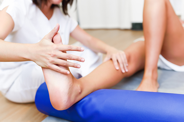
Physical Therapy
Physiotherapy is a key component of pain management and recovery after ACL surgery. Rehabilitation typically begins soon after the procedure, with early focus on reducing swelling, restoring knee extension, and activating the quadriceps muscles. As recovery progresses, exercises are adjusted to improve strength, balance, and neuromuscular control, supporting a smoother return to daily activities and, when appropriate, sports.
Are there some possible complications after an ACL surgery?

Infection
Although the incidence of infection after arthroscopic ACL reconstruction surgery is very low, there have been reports of deaths linked to bacterial infection from allograft tissue. This is due to improper procurement and sterilization techniques.
Stiffness
Stiffness is the feeling of difficulty in motion or the apparent loss of range of motion. It is sometimes accompanied by pain or swelling. Some patients have reported a loss of motion or stiffness in the knee after surgery.
Extensor mechanism failure
Rupture of the patellar tendon (patellar tendon autograft) or patella fracture (patellar tendon or quadriceps tendon autografts) may occur because of weakening at the site of graft harvest.
Growth plate injury
This is particularly applicable to young children or adolescents with ACL tears. Early ACL reconstruction creates a possible risk of growth plate injury, which can cause bone growth problems. The ACL surgery can be delayed until the child is closer to skeletal maturity. Alternatively, the technique of ACL reconstruction may be modified to lower the risk of growth plate injury.
Kneecap pain
Lastly, postoperative anterior knee pain is especially common after patellar tendon autograft ACL reconstruction surgery. The incidence of pain behind the kneecap varies widely in studies, while the incidence of kneeling pain is usually found to be higher after patellar tendon autograft ACL reconstruction.
Frequently Asked Questions About ACL Surgery in Singapore
How long does it take before I can return to work after ACL surgery?
The timing depends largely on the type of work you do. Patients in desk-based roles may return sooner, while those with physically demanding jobs often need more time before resuming full duties. Your recovery progress and functional stability will guide this decision rather than a fixed timeline.
Will an ACL tear increase my risk of knee arthritis later in life?
An ACL injury may increase the long-term risk of knee degeneration, particularly if there are associated meniscus or cartilage injuries. Appropriate treatment, rehabilitation, and activity modification can help reduce this risk and support long-term joint health.
How do I choose the right orthopaedic specialist for ACL surgery?
It helps to look for an orthopaedic specialist who has experience managing ACL injuries and can clearly explain both surgical and non-surgical options. A thorough consultation should address your activity level, recovery goals, and long-term knee health, allowing you to make an informed decision.
Is ACL surgery usually covered by insurance in Singapore?
Insurance coverage depends on the type of policy you hold and its specific terms. Some plans may cover hospitalisation, surgical fees, and post-surgery rehabilitation, while others may have exclusions or claim limits. At Specialist Orthopaedic Centre, an orthopaedic clinic at Mount Elizabeth, Novena, we work with a panel of recognised insurance providers in Singapore. It is advisable to check directly with your insurer and our team to understand your coverage before proceeding under our services.
Can ACL surgery be performed alongside treatment for other knee or traumatic injuries?
Yes. When an ACL tear occurs alongside fractures or injuries to other knee ligaments, surgical planning may be coordinated to address all affected structures. In such cases, elements of trauma surgery may be incorporated to stabilise the knee and support overall recovery.